Airway obstruction?
On your first time leading the ICU night shift in a District General Hospital, an urgent bleep call rushes you to ED Resus. A concerned Emergency Physician briefs you about his patient, a 49 y/o female awaiting elective surgical review for a multinodular goitre with retrosternal extension. The diagnosis was done a month ago by CT imaging and has not caused symptoms until now. The patient lives a fully independent life, is a non-smoker and does not use any drugs (licit or illicit).
She’s been brought to hospital by ambulance with a 1-day history of sudden-onset SoB, desaturating to 76% on room air. On arrival to Resus RR >40, SpO2 89% on 15L O2, unable to complete full sentences. There is no stridor or hoarseness of voice and no wheeze. The patient looks distressed and is evidently struggling to breathe. Despite sinus tachycardia, the BP is normal and the peripheral perfusion is adequate. A chest XR shows:
Severe tracheal deviation to the right. There is no evidence of atelectasis, consolidation, effusion or pneumothorax.
The ED doctor has decided the patient needs intubation to manage the airway obstruction and has contacted the Anaesthetics team who is on board with the plan and already assessing the patient for RSI. In the meantime, you give the details to the on-call ICU consultant who agrees with the admission yet is curious about the lack of stridor.
The patient is successfully intubated in Resus by the Anaesthetics team and subsequently connected to a mechanical ventilator. However, a couple of minutes after the intubation and despite adequate bilateral air entry, the SpO2 remains 91%. A control chest XR shows the tube adequately placed in the trachea and the deviation corrected.
Tracheal tube sitting above the bifurcation. The tracheal curve is now corrected, yet the trachea is still deviated to the right.
Still in Resus, an arterial line is placed for BP monitoring and she remains haemodynamically stable. Unable to optimise the SpO2 in spite of FiO2 100% and suspecting a PE, the patient is taken for a pulmonary angiography CT scan. While the images are still loading on the electronic system the patient arrives at ICU, where she suddenly deteriorates. She is on sinus tachycardia at 105bpm and the BP is plummeting to 69/45. The SpO2 remains around 90%.
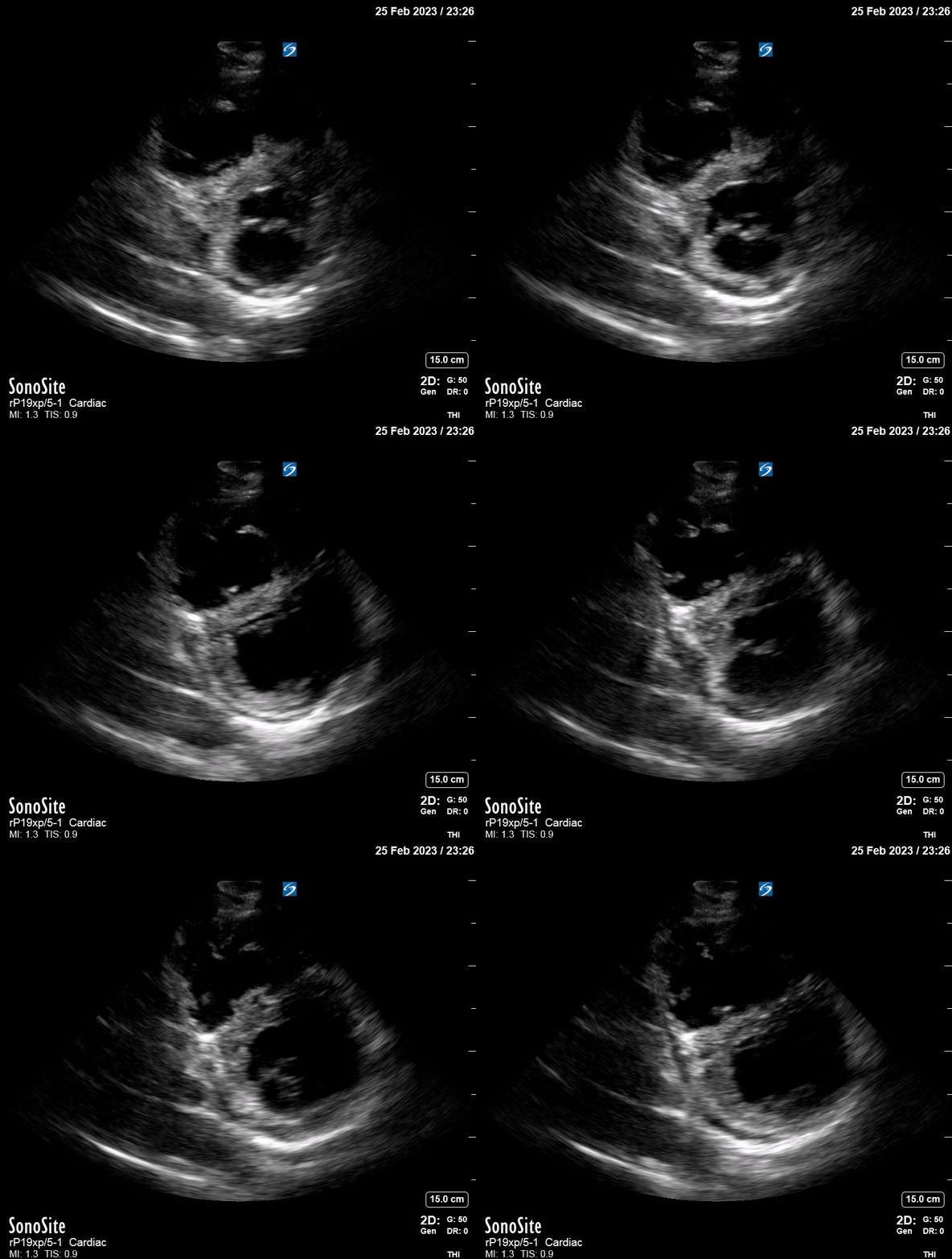
A push-dose of adrenaline (1 ml of 10ug/ml dilution) momentarily recovers the BP while the nursing team prepares an adrenaline infusion. Simultaneously, a critical care bed-side echo shows the following images:


The ICU consultant on-call has had a chance to look at the CT images and rings back to alert you about the presence of a massive bilateral PE. Upon discussion of the echo showing significant RV dilation with a collapsing septum, and the obvious circulatory shock the decision is made to provide emergency thrombolysis. The patient receives a bolus of Alteplase 50mg and a heparin infusion is started
CTPA demonstrated massive bilateral PE. Notice the opacified (darker) areas in both branches of the pulmonary artery.
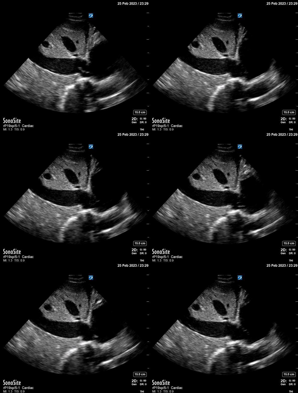
Within 30 minutes the haemodynamic instability has resolved, the adrenaline requirement is decreasing and the sats are back to 98%. Four hours later the RV strain is improving, the collapse of the septum resolving and the IVC shows a normal diameter with inspiratory variation.
A couple of days later the patient is off vasopressor support, has been extubated, is neurologically intact and able to walk. She is discharged home a week after.
Reflection
Although in hindsight the absence of stridor appears obvious and the decision to intubate questionable, on presentation the mutual agreement was to proceed this way. This highlights how fixation errors can be contagious and mislead you towards wrong management decisions.
In this case, the increase in intrathoracic pressure driven by positive pressure ventilation is likely to have worsened the obstructive shock and caused the marked haemodynamic deterioration.
In sum, this serves as evidence of the value of point-of-care ultrasound in the approach to acute dyspnoea. If possible, echo first!


